Important: This guide explains visual simulation. It does not diagnose a condition or recommend treatment.
Key takeaways
- A front-facing photo can explore visible width, length, shade and symmetry.
- It cannot show bone, roots, bite, gums or whether an implant is appropriate.
- Keep the preview as a preference reference for a proper examination and imaging review.
What a photo preview can show
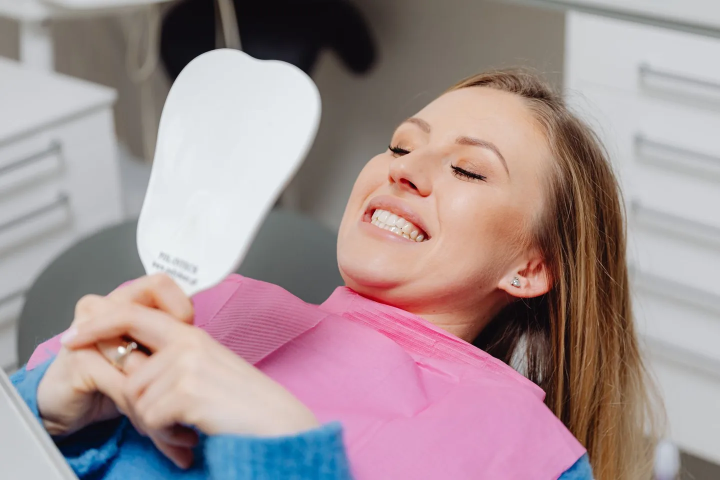
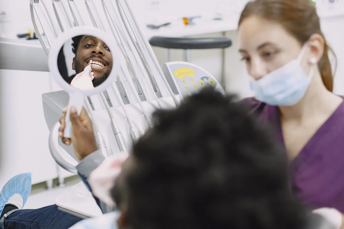
A smile preview can place a visible tooth shape into a front-facing gap and help you explore apparent width, length, shade and symmetry. This can make it easier to explain what feels natural to you.
What it cannot show
A photograph cannot confirm the condition underneath the visible gap.
- Available bone volume and density
- Root position of neighbouring teeth
- Bite and functional loading
- Gum thickness and expected tissue shape
- Whether an implant, bridge or another option is appropriate
Use the result as a conversation starter
Save the image as a preference reference. A dentist will still need appropriate examination and imaging before discussing realistic treatment options.
Why a front tooth needs more than a matching white shape
A believable visible replacement relates to the neighbouring tooth, gum edge, lip line and the way light crosses the smile. The apparent space in a flat photograph may also be distorted by camera angle. This is why a preview should be judged as an overall face image, not used as a measurement.
For a single front space, try a small range rather than one perfect-looking answer. A slightly warmer shade or a less symmetrical edge can sometimes blend more naturally than a mathematically identical copy.
What a clinical assessment adds
A clinician may review the mouth, gums, bite, neighbouring teeth and three-dimensional imaging before discussing implant, bridge or other restorative options. The sequence, healing time and temporary appearance may matter just as much as the final visible tooth.
If treatment may involve travel, ask what information can be assessed remotely and what can only be confirmed in person. Keep the visual preview separate from the provisional clinical plan, quotation and schedule so that one attractive image does not hide important uncertainty.
- Health and shape of the gum tissue
- Available bone and important anatomy
- Space between roots and the opposing bite
- Temporary replacement during treatment
- Material, shade and maintenance plan
Frequently asked questions
Can I use a missing tooth simulator for an implant?
You can explore the visible aesthetic direction, but implant suitability and position require examination and appropriate imaging.
Does the preview measure the gap accurately?
No. Camera distance, lens distortion and head angle change apparent dimensions, so the image should not be used for clinical measurement.
What should I send for a professional case review?
Follow the provider's instructions. A useful review commonly needs more than a smile photo and may require dental images, scans and health information.